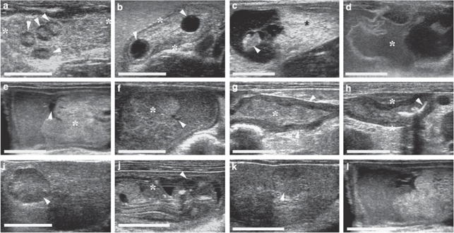
Figure 2. Ultrasonographic detection of embryonic resorption.
(a) In a preimplantative stage up to day 5 of gestation, the number of corpora lutea (white arrowheads), and hence the number of estimated embryos, can be counted. (b) Periimplantative (from days 6 to 10 of gestation), when embryonic vesicles can be seen but not yet a live embryo, the difference between the number of embryonic vesicles (white arrowheads) in the lumen of the uterus (*) and corpora lutea indicates early embryonic loss. (c–h) The direct ultrasonographic confirmation of postimplantative embryonic resorption can be carried out from day 11 of gestation onwards and typical stages of the course of embryonic resorptions can be visualized: (c) Loss of integrity: dead embryo (white arrowhead) but still intact placenta (*) and extraembryonic structures. (d) Embryo not detectable anymore and loss of integrity of extraembryonic structures (*). (e, f) Only small amounts of fluid remain (white arrowheads), decrease in size and compaction of resorption site, placental tissue (*) as the largest tissue part to be resorbed remains the longest. (g) Only undefined firm tissue (*) structures are left in the uterus (white arrowhead). (h) Last detectable stage of embryonic resorption: Scars (white arrowheads) from implantation in the endometrium of a uteruine loop (*). (i–l) Partial regression of corpora lutea according to partial embryonic resorptions: (i) active corpus luteum (white arrowhead) and (j) live embryo (eye (white arrowhead), heart (*)), (k) corpus luteum in regression (white arrowhead) and (l) resorption site. The white size bar indicates 10 mm.

