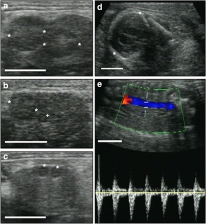
Figure 4. Ultrasonographic diagnosis of prepartum ovulation in free-ranging individuals.
(a) Section of left ovary with two corpora lutea of pregnancy (*). (b) Different section of left ovary with two different types of corpora lutea, one corpus luteum of pregnancy (*) 'belonging' to the fully developed first pregnancy, one small 'new' corpus luteum (+) from the second ovulation before parturition. (c) Section of right ovary with two small 'new' corpora lutea (*). (d) Section of the head (*) of a full-term fetus. (e) Doppler ultrasonogram indicating the heartbeat and liveliness of the full-term fetus and hence the demonstration of evidence for ovulations during an intact pregnancy in a free-ranging female. The white size bar indicates 10 mm.

