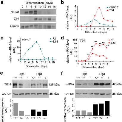
Figure 3. Hand1 regulates vascular differentiation in embryoid bodies.
Tβ4 and Hand1 levels were visualized by northern blotting (a) and quantified relative to Gapdh levels (b). Real-time qRT–PCR was used to measure Hand1 (c) or Tβ4 expression (d) during a time course of differentiation in wild-type (R1) and Hand1-null (6.13) EBs. Tβ4 levels are significantly downregulated in a Hand1-null background (day 6, *P ≤ 0.05; days 8–16, **P ≤ 0.01; Student's t-test) (d). Western blot analysis showed that vascular markers, both endothelial (e) and smooth muscle (f), are diminished in Hand1-null embryoid bodies (EBs), as quantified by scanning densitometry, normalized against GAPDH. Expression levels of these markers, TIE-2 (e) and α-SMA (f), are restored by supplementing the culture medium with synthetic TB4 (+Tβ4). Error bars in c and d represent s.e.m., where n = mean of three samples per time point. AU, arbitrary units.

