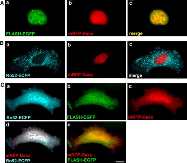
Fig. 7.
Subcellular localization of mRFP-Daxx in HT1080 cells expressing Ro52-ECFP and/or FLASH-EGFP. HT1080 cells were transfected to express: A FLASH-EGFP and mRFP-Daxx, B Ro52-ECFP and mRFP-Daxx, C Ro52-ECFP, FLASH-EGFP, and mRFPDaxx. The cells were fixed in a 4% paraformaldehyde solution and analyzed by fluorescence microscopy. The localization of Ro52-ECFP is shown by the cyan fluorescence of ECFP. The localization of FLASH-EGFP is shown by the green fluorescence of EGFP. The localization of mRFP-Daxx is shown by the red fluorescence of mRFP. The merged images are shown in A-c, B-c, C-d, and e as indicated. A scale bar indicates 10 μm

