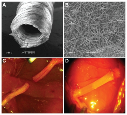
Figure 12.
Experimental model. Scanning electron microscopy images of the electrospun poly-D,L-lactic acid/polycaprolactone nerve guide conduit (A) and magnified details of the tube wall (B) microfibers and nanofibers range in diameter from approximately 280 nm to 8 μm. The nonwoven fibrous microstructure is characterized by small pores (700 nm) and large pores (20 μm). C) Micrograph of sham-operated rat sciatic nerve (experimental Group 1). D) Micrograph of prosthesis implanted, filled with saline solution, and sutured to the transected nerve (experimental Group 3).

