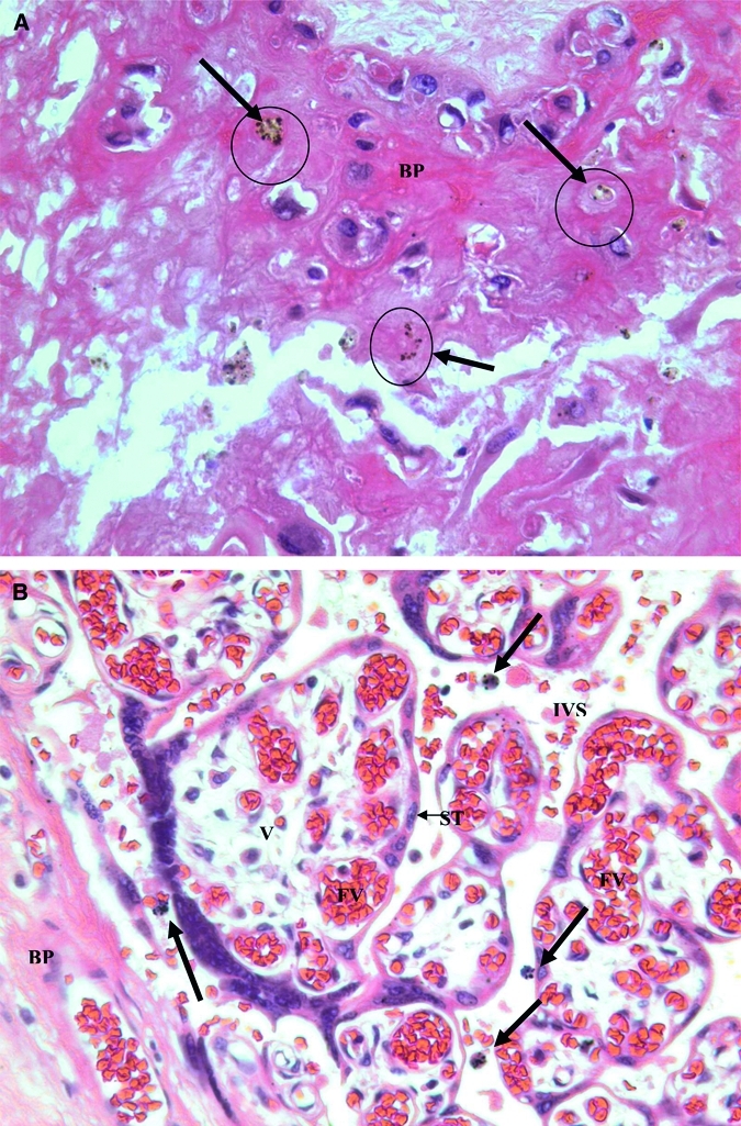
Figure 1.

A, Hemozoin deposition (Hz) in a basal plate (BP) of a placenta with evidence of past malaria infection. Malaria pigment or hemozoin appears as brown deposits (arrows). Pigment was confirmed as hemozoin by staining with Prussian blue. The pattern suggests that hemozoin was left in the fibrin of the basal plate after macrophage degeneration. B, Hemozoin deposition in maternal macrophages in the intervillous space. Four pigmented maternal monocytes (bold arrows) are in the intervillous space near the basal plate. BP = basal plate; IVS = intervillous space; V = fetal villi in cross section; ST = syncytiotrophoblast cells; FV = fetal vessels. (Original magnification × 400.)
