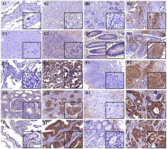
Figure 6.
HAGE protein expression in multiple cancer tissue microarrays and patient-matched normal tissues determined by immunohistochemistry. Immunohistochemical staining demonstrated the in vivo expression of HAGE protein at a low level in bladder transitional cell carcinoma (A2) and breast invasive ductal carcinoma (B2); at an intermediate level in astrocytoma (C2), colon adenocarcinoma (D2) and lung squamous cell carcinoma (E2); at a high level in esophagus small cell carcinoma (F2), kidney clear cell carcinoma (G2), hepatocellular carcinoma (H2), small intestine papillary adenocarcinoma (I2) and stomach adenocarcinoma (J2) but not in their respective matched normal tissues (A1, B1, C1, D1, E1, F1, G1, H1, I1 and J1). Objective magnification: 40x (inset 100x).

