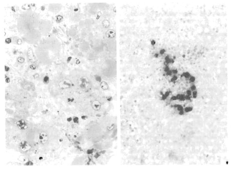
Fig 2.

Adenovirus hepatitis, Left, Case 1 (autopsy), infected nuclei are larger than uninfected ones. Nuclear “inclusions” consist of a patchwork of waxy-appearing dense material with unstained clear zones (1 μm) (polychrome, original magnification × 330). Right, Case 1 (biopsy specimen) frozen section of liver with a granuloma. Section has been stained with antiadenovirus monoclonal antibody, revealing dense reaction product in enlarged hepatocytes (amino-ethylcarbazol, original magnification × 97).
