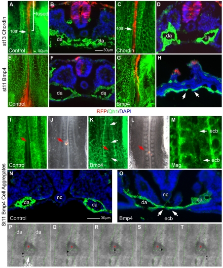
Fig. 5.
Misexpression of Chrd and BMP4 is sufficient to regulate dorsal aortae fusion. Whole-mount and sectioned embryos imaged for direct RFP fluorescence (red), endothelial-specific Qh1 antibody (green) and DAPI nuclear counterstain (blue) in sections. (A) Rfp-electroporated control embryo at fusion stage (stage 13) showing fused dorsal aortae at the 10th somite position (n=11). (B) Transverse section of A, showing normally fused dorsal aortae below RFP-expressing tissues. (C) Chrd+Rfp electroporated embryo showing successful inhibition of dorsal aortae fusion in 84% of embryos (n=13). (D) Section of C showing that bilateral dorsal aortae remained unfused. (E,F) As in A,B but pre-fusion-stage control embryo (stage 11) showing unfused bilateral dorsal aortae (n=11). (G,H) As in E,F but Bmp4+Rfp electroporated embryo showing midline vessels and fused aortae-like vessels (arrows). Midline vessels occurred in all BMP4-expressing embryos (n=20). (I) Pre-fusion stage 11 control embryo with implanted RFP-expressing cell aggregates (red arrow) showing bilateral dorsal aortae (n=11). (J) As in I but a composite of RFP and bright-field images visualizing RFP-expressing cell aggregates (arrow). (K) As in I but BMP4-expressing cell aggregates (red arrow) exhibit several fused regions of the dorsal aortae (white arrows, occurring in nine out of 17 embryo). (L) Composite of RFP and bright-field images of K. Note the BMP4+RFP expressing cell aggregates between two fused regions of the dorsal aortae. (M) Magnified view of fused regions (white arrows) of the dorsal aortae in L,K. (N) Transverse section of control pre-fusion-stage embryo with bilateral dorsal aortae near the cell aggregates (not in view). (O) As in N but with BMP-expressing cell aggregate implantation (not in view). (P-T) Sequence of still images from Movie S1 in the supplementary material showing path of single blood cells (circles/arrows) moving through an endothelial cell bridge between the aortae induced by BMP4-misexpression (red asterisks). Dorsal aortae and endothelial cell bridge are outlined by green dots. ecb, endothelial cell bridge; da, dorsal aortae; nc, notochord.

