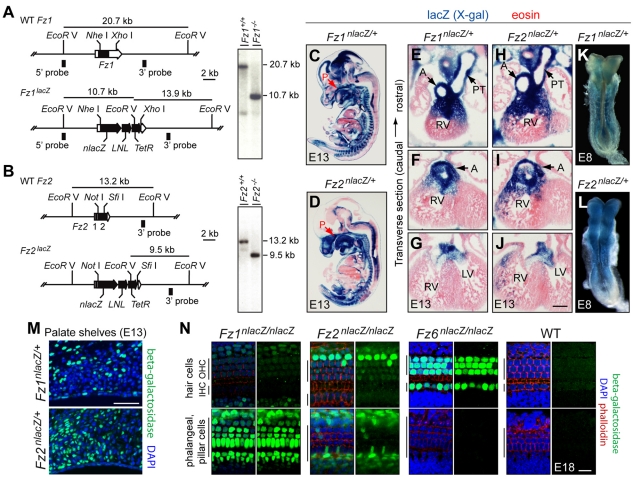
Fig. 1.
Targeted mutation of Fz1 and Fz2 in mouse and expression of Fz1nlacZ and Fz2nlacZ. (A,B) Fz1 and Fz2 knockout/nlacZ knock-in strategy. Wild-type (WT) Fz1 and Fz2 genes (upper maps), gene knockout/nlacZ knock-in alleles (lower maps), and genomic Southern blots (right). Southern blot probes and fragment sizes for EcoRV digests are shown. LNL, PGK-neo cassette flanked by loxP sites, later excised by germline Cre-mediated recombination. TetR, tetracycline resistance gene. (C-J) X-gal staining (blue) at E13 shows distinctive patterns of Fz1nlacZ and Fz2nlacZ expression, including in the developing palate epithelium and mesenchyme (C,D), and at the base of the cardiac outflow tract and the developing aorta and pulmonary trunk (E-J). P, palate; A, aorta; PT, pulmonary trunk; LV, left ventricle; RV, right ventricle. (K,L) X-gal staining (blue) of Fz1nlacZ/+ and Fz2nlacZ/+ E8 embryos shows Fz1 expression principally in the somites and surrounding mesenchyme, and widespread Fz2 expression, including in the lips of the open neural tube. (M) Anti-β-gal immunostaining (green) reveals Fz1nlacZ and Fz2nlacZ expression on the medial walls of the palate shelves at E13. Counterstained with DAPI (blue). (N) Anti-β-gal immunostaining (green) reveals Fz1nlacZ, Fz2nlacZ and Fz6nlacZ expression in the organ of Corti at E18; a wild-type control is shown on the right. Counterstained with DAPI (blue) and phalloidin (red) in each left-hand panel. The upper and lower rows of images show optical sections at the level of the hair cell nuclei and the underlying phalangeal and pillar cell nuclei, respectively. IHC, inner hair cells; OHC, outer hair cells. Scale bars: in J, 250 μm for E-J; 100 μm in M; 20 μm in N.

