Fig. 7.
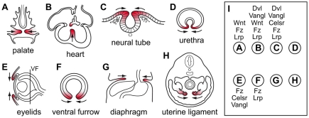
Diverse tissue closure processes in mammalian development, and the involvement of PCP and canonical Wnt signaling genes. (A-H) Eight tissue closure processes are shown. The plane of section is shown in E for the ventral furrow (VF) in the eye (F). The closing tissue is highlighted in red, and arrows indicate the direction of movement. (I) The general classes of PCP and canonical Wnt signaling genes that are involved in each closure process depicted in A-H are listed adjacent to the corresponding letters; in some cases, defects are only seen when more than one redundant or partially redundant gene is mutated.

