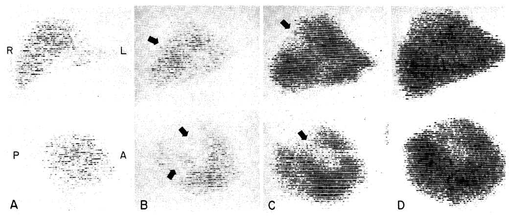
Fig. 5.
Post-transplantation liver scans in Case 1, performed with technetium-99m sulfide. For each examination an anteroposterior view is shown above and a lateral view below. A, 17 days; there are no defects B, 29 days; large nonvisualizing areas are evident (arrows). C, 32 days; 48 hours after debridement of the affected necrotic areas. D, 78 days; showing extensive regeneration into the previous defects.

