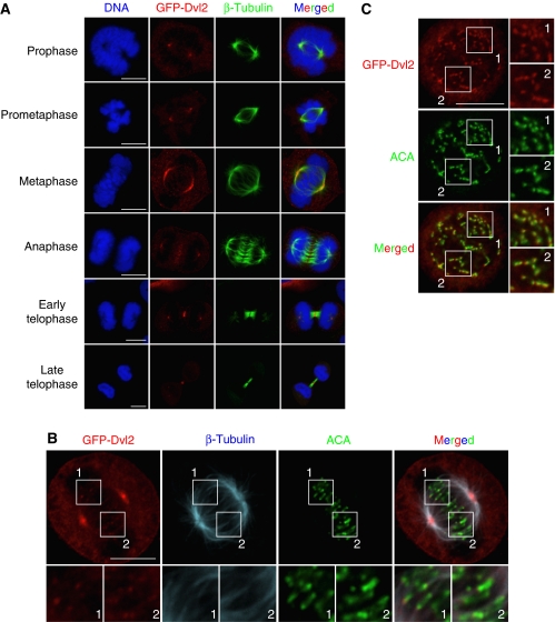
Figure 1.
Subcellular localization of Dvl2 in mitosis. (A) HeLaS3 cells transiently expressing GFP-Dvl2 were stained for GFP-Dvl2 (red) or β-tubulin (green). DNA (blue) was stained with PI. (B) HeLaS3 cells expressing GFP-Dvl2 at metaphase were stained for β-tubulin (blue), ACA (a KT maker, green), or GFP-Dvl2 (red). (C) HeLaS3 cells expressing GFP-Dvl2 were treated with 200 ng/ml nocodazole for 1 h to disrupt MTs. Cells were stained for GFP-Dvl2 (red) or ACA (green). The region in the white box is shown enlarged. Scale bars in (A), (B), and (C), 10 μm.

