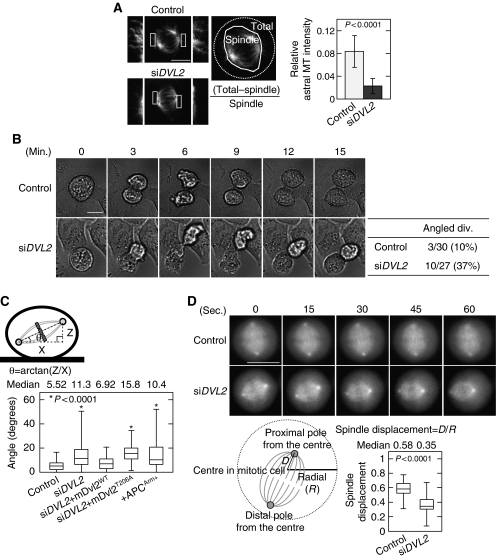
Figure 3.
Dvl2 regulates spindle orientation. (A) β-Tubulin was stained in control or Dvl2-depleted HeLaS3 cells, and the average relative astral MT intensity was calculated. The intensities of the total MT area and the total spindle area were measured and the relative astral MT intensity is expressed as the ratio of [(intensity of the total MT area)−(intensity of the spindle MT area)]/(intensity of the spindle MT area). Border between astral MT and spindle MT was determined by spindle morphology and intensity threshold. The regions in white boxes are shown enlarged. (B) Images of the mitotic control or Dvl2-depleted HeLaS3 cells were acquired every 3 min for 24 h. Mitotic cells were identified from the movie pictures (Supplementary Movies 1 and 2), and then counted as ‘plane division' (both daughter cells remained attached to the substratum after cell division) or ‘angled division' (one of two daughter cells failed to gain adhesion). (C) Top panels, scheme describing spindle angle (θ) measurement. Bottom panels, spindle angles were measured in HeLaS3 cells transfected with the indicated siRNA (si) or plasmids (+). siDVL2+mDvl2WT and siDVL2+mDvl2T206A indicates Dvl2-depleted HeLaS3 cells expressing GFP-mDvl2WT and GFP-mDvl2T206A, respectively. (D) Top panels, for visualizing the spindle in control or Dvl2-depleted cells, HeLa cells stably expressing GFP-EB3 was transfected with each siRNA. At 72 h after transfection, imaging was started. Images of the mitotic control and Dvl2-depleted HeLaS3 cells were acquired every 3 s (Supplementary Movies 3 and 4, respectively). Snap shots are shown at 15 s each. Bottom panels, the radial (R) of a metaphase cell and the distance (D) from the centre to the proximal spindle pole in a metaphase cell were measured, and spindle displacement was expressed as a ratio of D to R (n=65). Thirteen cells were captured by time-lapse imaging, and five snap shots were selected at 15 s each from one cell. Scale bars in (A), (B), and (D), 10 μm.

