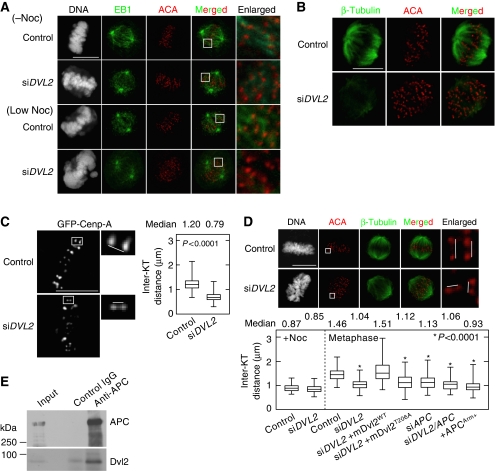
Figure 4.
Dvl2 regulates MT-KT attachment. (A) Control or Dvl2-depleted HeLaS3 cells were treated with or without a low concentration of nocodazole (12 ng/ml) in the presence of 10 μM MG132 (to arrest cells at metaphase), and the cells were stained with PI (grey), anti-EB1 (green; an MT plus-end marker) antibody, and ACA (red). The regions in white boxes are shown enlarged. (B) Control or Dvl2-depleted HeLaS3 cells were treated with a low concentration of nocodazole (12 ng/ml) in the presence of 10 μM MG132 (to arrest cells at metaphase), and the cells were stained with anti-β-tubulin (green) antibody (to observe K-fibres) and ACA (red). (C) Control or Dvl2-depleted living HeLa cells expressing GFP-Cenp-A were arrested at metaphase by treatment with 10 μM MG132 for 1 h. Images of cells were captured every 3 s for 3 min. Each KT pair was identified from the movie pictures and the inter-KT distance was measured (control, n=627 KT pairs; siDVL2, n=586 KT pairs). (D) The inter-KT distance of fixed HeLaS3 cells transfected with the indicated siRNAs (si) or plasmids (+) was measured at metaphase (n=100 KT pairs). Each KT pair was carefully identified from Z-stack images from 0.2 μm-thick sections of a cell. siDVL2+mDvl2WT and siDVL2+mDvl2T206A indicate Dvl2-depleted HeLaS3 cells expressing GFP-mDvl2WT and GFP-mDvl2T206A, respectively. (E) Lysates from mitotic HeLaS3 cells were immunoprecipitated with anti-APC antibody, and immunoprecipitates were probed with anti-APC and anti-Dvl2 antibody. Scale bars in (A), (B), and (C), 10 μm.

