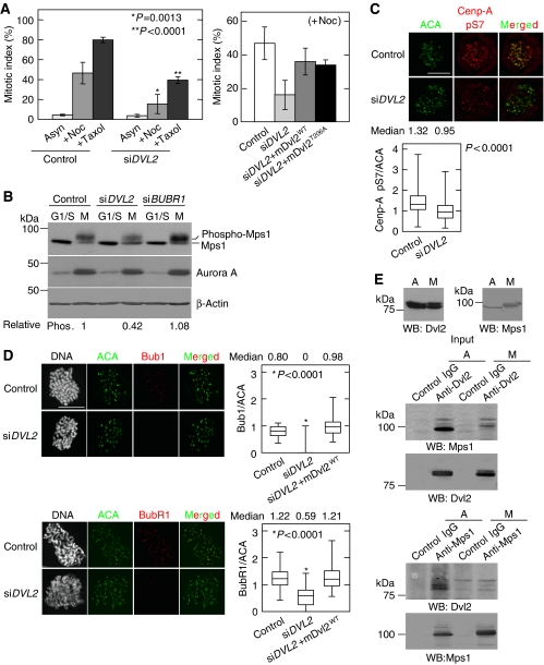
Figure 5.
Dvl2 is involved in the SAC. (A) Left panel, control or Dvl2-depleted HeLaS3 cells were treated with 100 ng/ml nocodazole or 10 nM taxol for 24 h and then stained with an anti-MPM2 antibody and PI. The ratios of mitotic cells (mitotic index) among >100 cells were calculated. Right panel, GFP-mDvl2WT or GFP-mDvl2T206A was expressed in Dvl2-depleted HeLaS3 cells treated with 100 ng/ml nocodazole, and the mitotic index was calculated after nocodazole treatment. (B) Control or Dvl2-depleted HeLaS3 cells were synchronized as shown in Supplementary Figure S2B. Lysates from G1/S phase (G1/S) or mitotic phase (M) cells were probed with the indicated antibodies. β-Actin was used as a loading control. (C) Control or Dvl2-depeleted HeLaS3 cells were treated with10 μM MG132 for 2 h, and then the cells were stained for ACA (green) and phospho-Ser7 of Cenp-A (red). The peaked intensities of Cenp-A pS7 and ACA were measured at each KT, and the ratio of the intensity of Cenp-A pS7 to ACA was calculated (control, n=141 KT pairs; siDVL2, n=240 KT pairs). (D) Control cells, Dvl2-depleted HeLaS3 cells, or Dvl2-depleted HeLaS3 cells expressing GFP-mDvl2 were treated with 200 ng/ml nocodazole for 1 h to activate SAC. After antibody reactions, DNA was stained with PI, and then mitotic cells were identified by condensed DNA. Peaked intensity of Bub1, BubR1, and ACA was measured at each KT in mitotic cells, and the ratio of the intensity of BubR1 or Bub1 to ACA was calculated (n=100 KT pairs). (E) Lysates from asynchronous (A) and thymidine-nocodazole blocked (M) HeLaS3 cells were immunoprecipitated with the indicated antibodies, and the immunoprecipitates were probed with anti-Dvl2 and anti-Mps1 antibodies. Scale bar in (C) and (D), 10 μm.

