Figure 1.
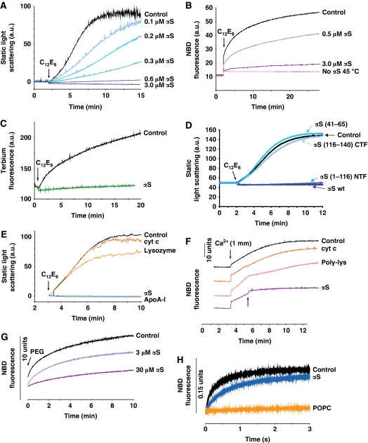
αS inhibits membrane fusion in vitro. (A) Fusion of DPPC-SUV was monitored by the increase in static light scattering upon addition of an aliquot of C12E8. Increasing amounts of αS inhibit membrane fusion (blue lines). Lipid concentration 600 μM, T=36°C. (B) Lipid-mixing assay of DPPC-SUV. αS inhibited fusion completely at lipid/αS=200 mole/mole (purple line). T=35°C. Pink line: no fusion occurred at 45°C, that is at temperature above Tm. (C) Contents-mixing assay carried out in the presence and absence of αS (lipid/αS=200 mole/mole, green line). T=30°C. (D) An N-terminal fragment mutant of αS, αS(1–116) (blue line), lacking the negatively charged C-terminal domain was capable of completely suppressing the fusion of DPPC-SUV, like wt-αS (dark blue line); whereas peptides comprised of the C-terminal fragment, αS(116–140), or the central domain of αS, αS(41–65), failed to inhibit fusion (blue lines). Lipid/protein=100 mole/mole. T=25°C. (E) Comparison of inhibition of membrane fusion by αS with cytochrome c, lysozyme and Apolipoprotein A-I (ApoA-I). For all proteins: lipid/protein=200 mole/mole. T=36°C. (F) Ca2+-induced fusion of POPS-SUV. Fusion was initiated by adding an aliquot of CaCl2 and monitored by the lipid-mixing assay. αS, added 2 min after the addition of Ca2+ (arrow), blocked fusion almost completely (lipid/αS=200 mole/mole). Control experiments: cytochrome c (lipid/protein=20 mole/mole) or poly-lysine (lipid/protein 200 mole/mole) was added instead of αS. T=25°C. (G) SUV of a mixture of lipids with reported optimal fusion potential (DOPC/DOPE/BBSM/cholesterol, 35:30:15:20 molar ratio) (Haque et al, 2001). Fusion was initiated by addition of 4% PEG and followed using the lipid-mixing assay. Total lipid concentration was 300 μM. When the experiment was repeated with αS, fusion was slowed. T=37°C. (H) Spontaneous rapid fusion of SUV composed of lipids with opposite charges (POPS-SUV and PC+-SUV). Lipid-mixing assay performed in stop-flow fluorimetry. Lipid concentration was 60 μM. αS (1.2 μM) inhibited the fusion. When POPS was replaced by POPC (uncharged) no fusion occurred, as expected. T=25°C.

