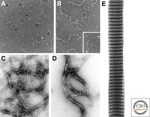
Figure 2.
Assembly of the nuclear lamins in vitro. Lamins self assemble to form dimers (A) which then join to form linear head-to-tail polymers (protofilaments) (B). Bar = 100 nm; electron micrograph of rotary shadowed chicken lamin B2. These protofilaments further assemble into “beaded” filaments or fibers (C) which in turn associate laterally into thicker fibers (D), and eventually into paracrystalline arrays (E); C,D are negatively stained electron microscope preparations. Reprinted from Stuurman et al. (1998) with permission from Elsevier.

