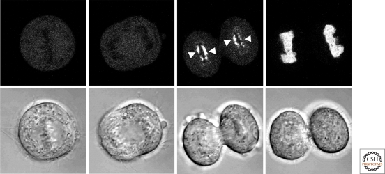
Figure 5.
Association of lamin A with chromosomes during mitosis. HeLa cells expressing GFP-lamin A were followed by time-lapse microscopy from the metaphase/anaphase transition (far left panels) into early G1 (far right panels). GFP-lamin A first associates with the core regions of chromosomes during telophase and spreads to cover the entire chromatin surface by early G1. DIC images of the same series are shown in the bottom row (Dechat et al. 2007).

