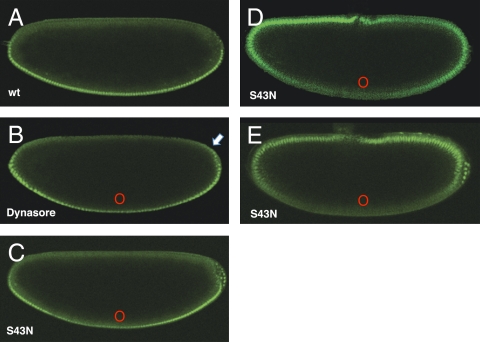
Fig. 3.
Inhibition of endocytosis ventrally reduces nuclear accumulation of Dorsal proximally and can alter the polarity of the Dorsal gradient. Live imaging of transgenic embryos expressing Dorsal-GFP at nuclear cycle 14. (A) Mock-injected control embryo expressing Dorsal-GFP showing the WT (wt) nuclear Dorsal gradient. (B) Embryo microinjected with Dynasore, a dynamin inhibitor, as a small bolus on the ventral midline in which nuclear accumulation of Dorsal is attenuated near the site of injection and slightly expanded dorsally (arrow). (C) Embryo injected with synthetic mRNA encoding Rab5S43N, a dominant negative form of Rab5 on the ventral midline. (D) Embryo injected as in C and allowed to develop for 40 min, illustrating a progressive shifting of nuclear translocation to the dorsal side of the embryo. (E) Rab5S43N ventrally injected embryo showing complete inversion of the Dorsal gradient. In all cases, ventral is down, dorsal is up, and red circles mark the site of injection of drug or synthetic mRNA.

