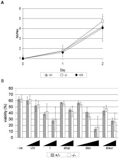
Figure 2. Normal proliferation and apoptosis of Anp32e−/− cells.
A. Growth curves of primary MEFs from Anp32e+/+, Anp32e+/− and Anp32e−/− mice (n = 2/genotype). Nt/N0, cell number at time point/cell number on day 0. B. Apoptosis of Anp32e+/− and Anp32e−/− thymocytes after 20 hours in culture response to (left to right): –ve, untreated controls, UV-irradiation (30 mJ/cm2 and 60mJ/cm2), γ-irradiation (1 Gy and 2 Gy), etoposide (1 µM and 3 µM), dexamethasone (3 nM and 10 nM), and staurosporine (1 µM and 3 µM). Results shown are mean % viable cells ± SD (n = 3/genotype).

