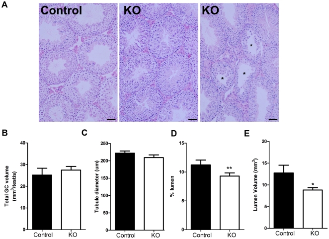
Figure 3. Histological comparison of adult SMARKO and control testes.
A. Most seminiferous tubules in SMARKO testes looked comparable to control testes at d100, however, a small proportion (10%) were abnormal with disturbed spermatogenesis (*). Lumens appeared smaller in many seminiferous tubules in SMARKO testes at d100, compared to controls. B. There was no significant change in the total germ cell volume in SMARKO testes at d100, compared to controls. C. There was no significant change in seminiferous tubule diameter in SMARKO testes at d100, compared to controls. The percentage (D) and volume (E) of seminiferous tubule lumen was significantly reduced at d100 in SMARKO testes compared to controls. Scale bars = 50 µm. Values are means ± S.E.M. (n = 4–6 mice), * p<0.05, ** p<0.01 compared to controls littermates.

