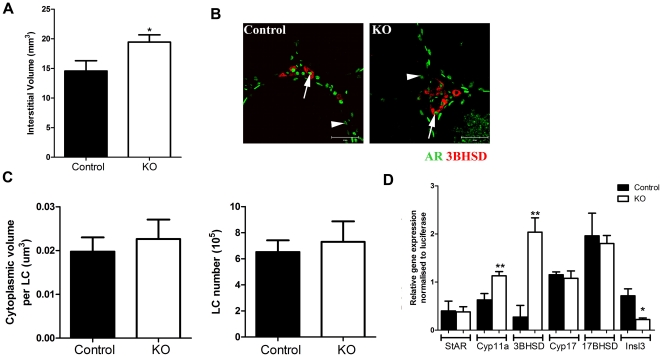
Figure 4. Evaluation of Leydig cell (LC) function in SMARKO testes.
A. There was a significant increase in interstitial volume in SMARKO adult testes. B. Immunostaining for 3βHSD (red) and AR (green) demonstrating that LCs (arrow) and SCs (arrowhead) express AR in both SMARKO and control testes. C. Quantification of LC size and number highlighting no significant change in either in SMARKO testes, compared to controls. D. Relative expression of steroidogenesis enzymes (StAR, cyp11a, 3βHSD, Cyp17 and 17βHSD) and Inls3 in d100 control and SMARKO testes. Values are mean ± SEM (n = 4–6 mice), ** p<0.01, compared to controls littermates. Scale bars = 50 µm.

