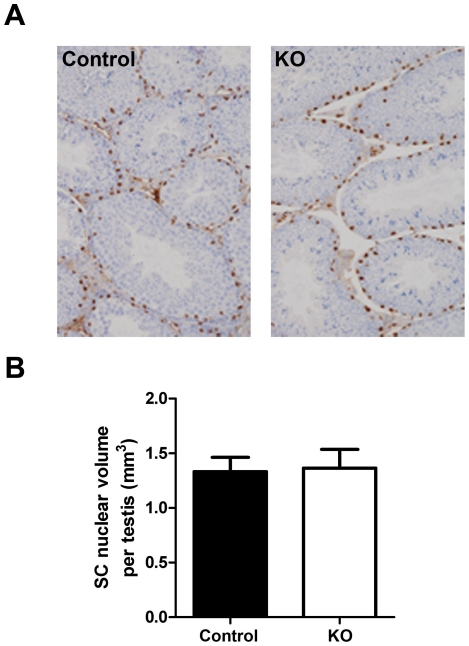
Figure 6. Evaluation of Sertoli cell (SC) function in SMARKO testes.
A. WT-1 staining (black) was used to identify SC nuclei and highlighted their basal location in SMARKO and control males. B. SC mean nuclear volume in SMARKO and control d100 testes. Values are means ± S.E.M. (n = 4–6 mice). Scale bars = 50 µm.

