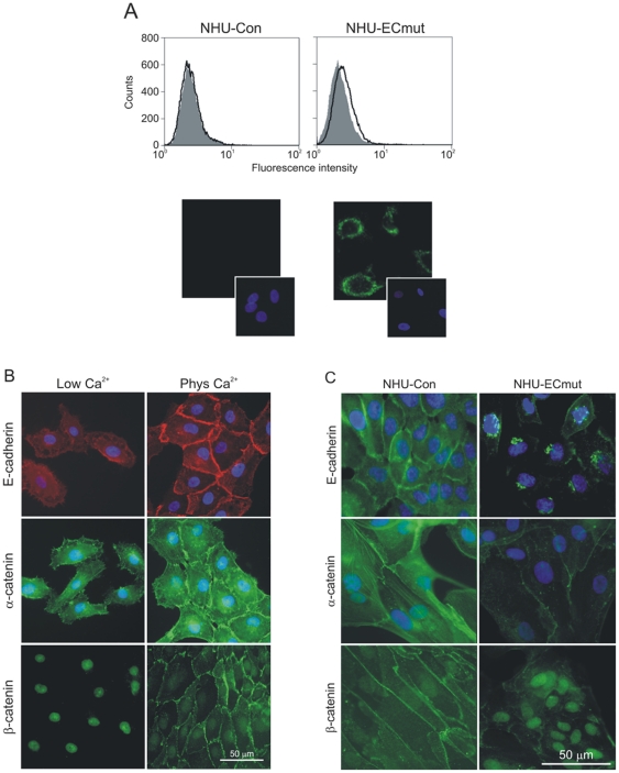
Figure 3. Effect of dominant-negative mutant E-cadherin on expression and localisation of endogenous E-cadherin and catenins in NHU cells.
(A) NHU cells expressing the H-2Kd-E-cad mutant (NHU-ECmut) and their isogenic controls (NHU-Con) were established by retrovirus transduction and expression of mutant E-cadherin was assessed by flow cytometry and immunofluorescence microscopy. For flow cytometry (upper panels), expression was analysed using FITC-conjugated anti-H-2Kd antibody (open histograms) alongside irrelevant isotype-matched control antibody (filled histograms). Histograms represent log10 fluorescence intensity in the FL-1 channel. For immunofluorescence microscopy (lower panels), expression was detected using anti-H-2Kd antibody followed by goat anti-mouse antibody conjugated with Alexa Fluor 488 (green). (B) Following initial seeding, NHU cells were cultured in medium containing low (0.09 mM) or physiological (2.0 mM) Ca2+ concentrations for 24 hours before expression of E-cadherin, α-catenin and β-catenin was assessed by microscopy using mouse (E-cadherin) and rabbit (catenins) antibodies, followed by goat antisera conjugated with Alexa Fluor 488 (green) or 594 (red). (C) NHU-Con and NHU-ECmut cells were cultured in medium containing physiological [Ca2+] and expression of E-cadherin, α-catenin and β-catenin was determined by labelling using primary antibodies above, followed by Alexa Fluor 488-conjugated antibody (green). In all immunofluorescence microscopy experiments, cell nuclei were visualised by labelling with Hoechst 33258 (blue).

