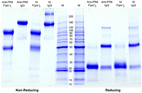
Figure 1. SDS-PAGE of IgG and F(ab')2 preparations.
Protein samples in SDS buffer were subjected to electrophoresis under non-reducing and reducing conditions through a 4–20% Tris-HEPES SDS-PAGE gel. The gel was stained with Coomassie blue G-250. Molecular mass markers (M) are indicated in kilodaltons. NI, non-immune. The minor band in the non-immune IgG preparation run under non-reducing conditions corresponds to IgG dimers which can sometimes form after freeze-drying. The results are typical of profiles from three separate SDS-PAGE analyses of these particular preparations and of many other analyses of colostrum-derived antibodies .

