Fig. 1.
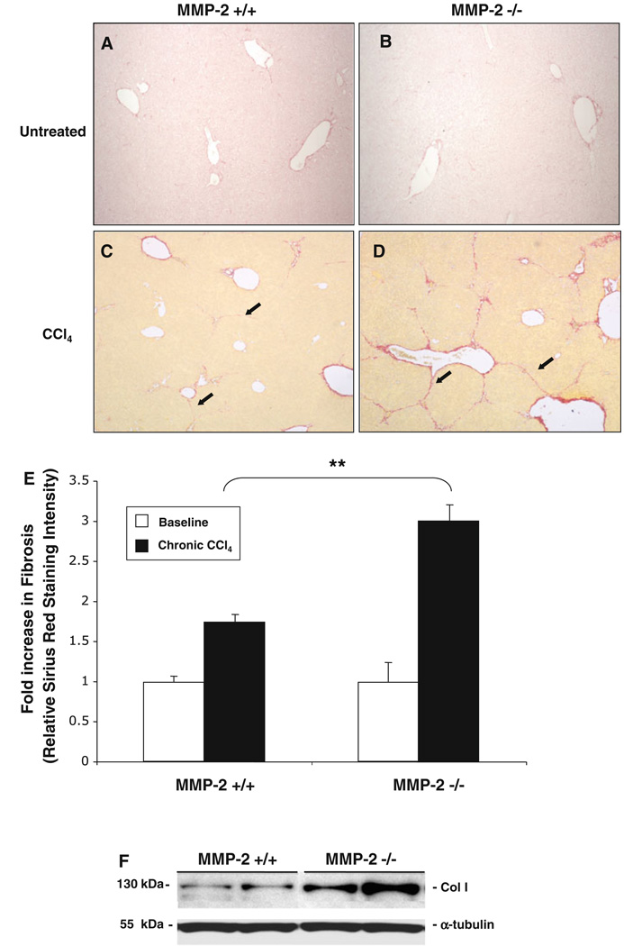
MMP-2−/− mice develop increased hepatic fibrosis in response to chronic liver injury. MMP-2+/+ and MMP-2−/− livers were harvested and sectioned, and degree of fibrosis assessed with Sirius Red staining at baseline and following chronic CCl4 administration. Percentage overall fibrosis was subsequently calculated via histomorphometric bioquant analysis of 36 images per animal (n = 4) in a blinded fashion. Representative baseline Sirius Red staining on MMP-2+/+ (a) and MMP-2−/− (b) livers and examples of bridging fibrosis after chronic CCl4 in MMP-2+/+ (c) and MMP-2−/− (d) mouse livers shown. Results of histomorphometric bioquant demonstrates a 1.7-and 3-fold increase in fibrosis in MMP-2+/+ and MMP-2−/− livers from untreated baseline, respectively. An almost twofold increase in fibrosis was observed in the MMP-2−/− livers as compared with the MMP-2+/+ livers after chronic CCl4 administration (e). Representative immunoblot using whole liver protein extracted from MMP-2+/+ and MMP-2−/− mice after chronic CCl4 administration confirms increase in collagen I expression (f). Original magnification 40×. Arrows point to areas of bridging fibrosis. Data represent means ± SEM; ** P < 0.001

