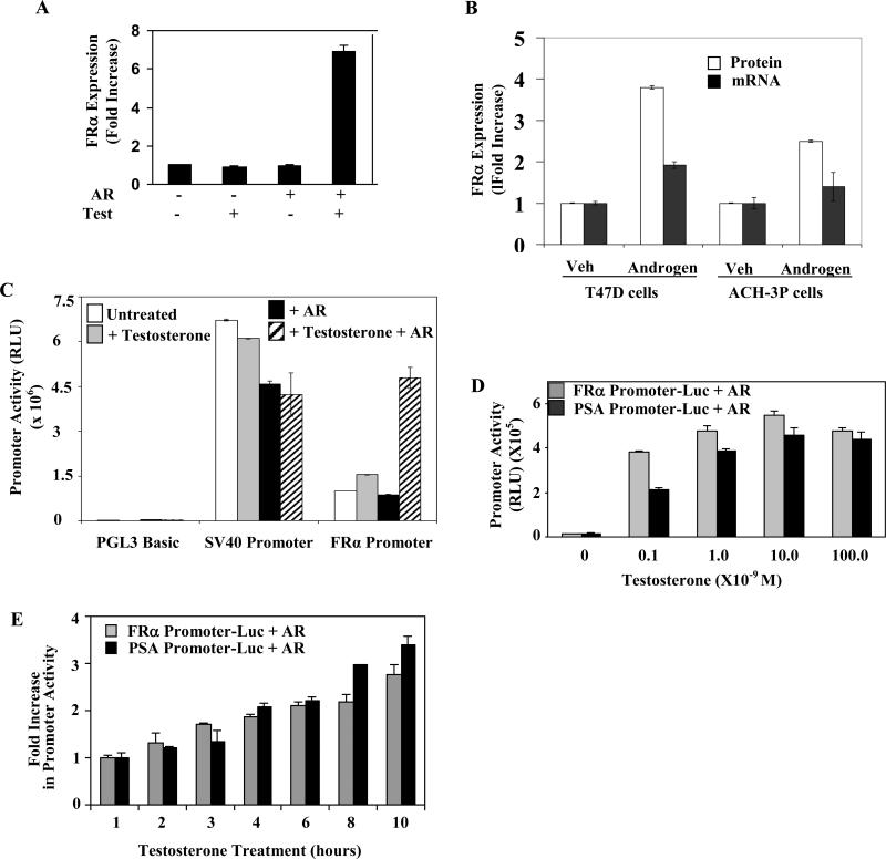
Figure 1. Specific regulation of FRα by androgen.
(A) HeLa cells transfected with AR expression plasmid were treated with 10 nM testosterone (Test) or vehicle for 3 days. FRα cell surface expression was quantified. (B) T47D cells or ACH-3P cells were treated with 10 nM R1881 or vehicle for 72h. FRα cell surface expression and the FRα mRNA were measured. (C) Hormone deplete HeLa cells were transfected with one of the following constructs: the FRα promoter Luc, SV40 promoter Luc or PGL3 Basic Luc (empty vector control plasmid). The cells were co-transfected with either the expression plasmid for AR or the vector control as well as the Renilla luciferase plasmid (transfection control) and either untreated or treated for 48h with testosterone (10 nM). The cells were then harvested to measure luciferase activity (D) Hormone deplete HeLa cells were transfected with the FRα promoter Luc or the PSA promoter Luc. The cells were co-transfected with either the expression plasmid for AR as well as the Renilla luciferase plasmid (transfection control) and either untreated or treated for 48h with different doses of testosterone. The cells were then harvested to measure luciferase activity. (E) Hormone deplete HeLa cells were transfected with the FRα promoter Luc or the PSA promoter Luc. The cells were co-transfected with either the expression plasmid for AR as well as the Renilla luciferase plasmid (transfection control). 48h post-transfection, the cells were either untreated or treated for different periods with testosterone (10 nM). The cells were then harvested to measure luciferase activity. P values for the differences noted in the text were < 0.001.

