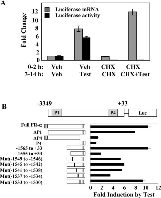
Figure 2. Direct regulation of FRα by androgen and mapping of the regulatory elements.
(A) HeLa cells were transfected with FRα (-1565 to +33) promoter-luciferase reporter and co-transfected with AR; 24h later, the cells were then treated with either 10 μM cycloheximide (CHX) or vehicle for 2 h followed by the introduction of testosterone (10 nM) or vehicle for further 12h. Total RNA was extracted for quantitation of luciferase mRNA by real-time reverse transcription-PCR. In a parallel experiment, the cells were harvested to measure the luciferase protein expression by measuring luciferase activity. (B) HeLa cells were transfected with the following promoter-luciferase reporter constructs: the full FRα promoter [FRα(-3394nt to +33nt)-luc]; the full FRα promoter with deletion of either the P1 promoter (ΔP1) [FRα(-3113nt to +33nt)-luc] or the P4 promoter (ΔP4) [FRα(-3394nt to +33nt, Δ-146 nt to -34nt)-luc]; the P4 promoter [FRα (-176 nt to +33nt)-luc]; the FRα promoter with 5’ deletions (-1565nt to +33nt and -1555nt to +33nt); the FRα promoter constructs with sequential 4 base pair mutations from -1549nt through -1530nt. The positions of the mutated nucleotides are indicated by the vertical black bars (not to scale). The cells were co-tranfected with AR followed by treatment with either testosterone (10 nM) or vehicle and harvested for luciferase assay 48h post-transfection. P values for the differences noted in the text were < 0.001.

