Figure 1.
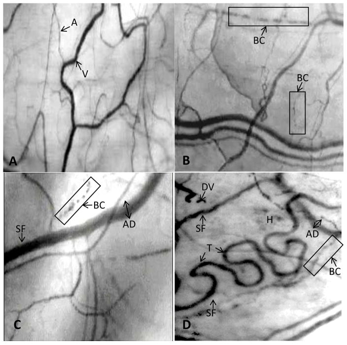
Figure 1A. A frame-captured image of the conjunctival microcirculation in a healthy non-SCA control subject [10, 11].
Optical magnification 4.5X; onscreen magnification 125X. This image illustrates a typical view of the conjunctival microcirculation in a healthy (non-SCA) control subject who has no history of any vascular disease. Note the even and orderly distribution of normal-sized arterioles, venules and capillaries in a richly vascularized network.
Abbreviations: A = arteriole; V = venule.
Figure 1B. A frame-captured image of the conjunctival microcirculation in a pediatric SCA patient (Patient #P-3; age 8y).
Optical magnification 4.5X; onscreen magnification 125X. The SI of this patient is 3 and the microvascular abnormalities include only sludged blood flow (vessel sludging), boxcar (trickled) blood flow and abnormal A:V ratio. Overall, the vasculopathy observed is mild.
Abbreviations: BC = boxcar (trickled) blood flow.
Figure 1C. A frame-captured image of the conjunctival microcirculation in another pediatric SCA patient (Patient #P-8; age 15y).
Optical magnification 4.5X; onscreen magnification 125X. Patient P-8 is 7 years older than the patient described in Figure 2. The microcirculation shows a greater level of vasculopathy, which includes abnormal vessel diameter, sludged blood flow, boxcar (trickled) blood flow, abnormal vessel distribution, hemosiderin deposit, and abnormal A:V ratio in this captured frame. The overall vasculopathy in this pediatric patient is severe, with an SI of 7 (compared with the SI of 3 in the pediatric patient described in Figure 2).
Abbreviations: SF = sludged blood flow [stop-and-go pattern of blood flow as evidenced by area(s) of darker or uneven coloration within the vessel]; BC = boxcar (trickled) blood flow; AD = abnormal diameter (wide).
Figure 1D. A frame-captured image of the conjunctival microcirculation in an adult SCA patient (Patient #A-7; age 58y).
Optical magnification 4.5X; onscreen magnification 125X. The microvascular abnormalities in this adult patient include abnormal vessel diameter, pronounced vessel tortuosity, abnormal vessel distribution, abnormal A:V ratio, sludged (trickled) blood flow, boxcar flow pattern, damaged vessel, and hemosiderin deposits.
Abbreviations: SF = sludged blood flow [stop-and-go pattern of blood flow as ecidenced by area(s) of darker or uneven coloration within the vessel]; BC = boxcar (trickled) blow flow; DV = damaged vessel; AD = abnormal diameter (wide); H = hemosiderin deposits; T = tortuosity.

