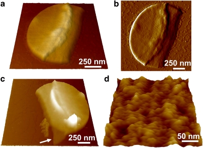
Figure 4. Sacculi do not show parallel bands.
(a, c, d) Height and (b) deflection images in air of peptidoglycan sacculi from L. lactis VES5748, gently broken by a french press. Images a and b show the outside surface of the same sacculus consisting of a double cell wall. Image c represents another sacculus in which the boarder exposes the inner surface of a single wall (arrow). (d) High-resolution view of the outer surface, similar morphology being observed on the inner surface.

