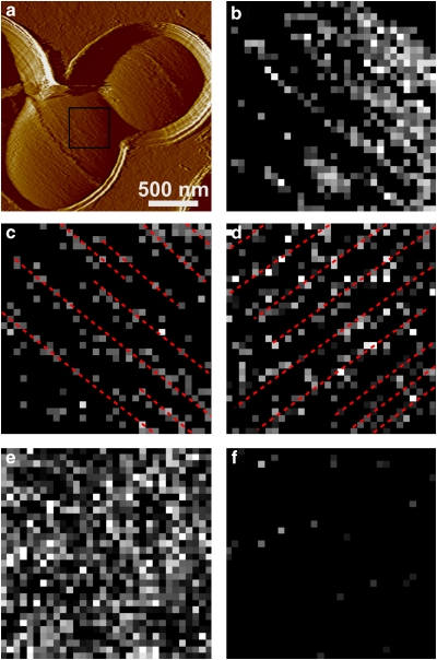
Figure 6. Peptidoglycan localizes as parallel lines on the surface of WPS− mutants.
(a) Deflection image of an L. lactis VES5748 WPS− mutant cell recorded with a silicon nitride tip. (b) Adhesion force map (400×400 nm) recorded with an LysM tip in the square area shown in the deflection image using a maximum applied force of 250 pN. (c–f) Adhesion force maps (500×500 nm) recorded with an LysM tip on another cell. Maps shown in c and d were recorded in the same area, except that the cell was rotated by 90°. Many of the detected molecules (bright pixels) were arranged as lines running parallel to the short cell axis (red lines). The map shown in e was obtained with a maximum applied force of 500 pN instead of 250 pN, whereas the map in f was obtained in a 10 μg ml−1 peptidoglycan solution. In addition to the data shown, similar results were obtained in nine different cells from at least five different cultures, using 10 different tips from at least five different batches.

