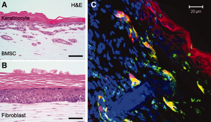
Figure 1.

Bone marrow-derived mesenchymal stem cells (BM-MSCs) in cutaneous regeneration. (A, B): Keratinocytes loaded on a collagen gel containing BM-MSCs formed rete ridge-like structure, whereas keratinocytes loaded on a collagen gel containing dermal fibroblasts did not. Images adapted from Aoki et al. [58]. Image courtesy of Molecular Biology of the Cell. (C): BM-MSCs (green) from GFP-expressing mice were injected around the excisional wound and applied on the wound bed in Matrigel in Balb/C mice. At day 7, some mesenchymal stem cells (yellow) expressed keratinocyte marker cytokeratins (red) and formed structures similar to those observed in the study of Aoki et al. (A). Abbreviations: BMSC, bone morrow-derived mesenchymal stem cells; H&E, hematoxylin and eosin stain.
