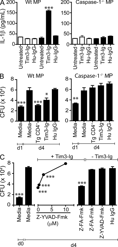
Figure 5.
Tim3–Gal9 interaction induces caspase-1–dependent secretion of IL-1β and Mtb killing. (A) Mtb-infected WT C57BL/6J macrophages (MP) or Caspase-1−/− macrophages were cultured alone or with 10 µg/ml of Tim3-Ig fusion protein or HuIgG (control). 24 h later, culture supernatants from triplicate wells were assayed for IL-1β. Open bars indicate uninfected macrophages, and closed bars indicate Mtb-infected macrophages. Data are representative of seven independent experiments. Error bars indicate mean ± SEM from three replicate cultures. ***, P < 0.001, one-way ANOVA compared with untreated macrophages alone. (B) WT C57BL/6J or caspase-1−/− macrophages were infected with H37Rv in parallel. On day 1, Tim3Tg CD4+ T cells, Tim3-Ig, or HuIgG (control) were added to the macrophages. CFUs were determined on day 1 and day 4 (d4) after infection. Data are representative of three independent experiments. Bars indicate mean ± SEM from three replicate cultures. (C) WT C57BL/6J macrophages infected with H37Rv was co-cultured with Tim3-Ig in the presence or absence of Z-YVAD-Fmk (caspase-1 inhibitor) titrated fivefold. CFUs were determined on day 0, 2 h after Mtb infection, and on day 4. Z-FA-Fmk, negative control peptide for caspase-1 inhibitor. HuIgG, control for Tim3-Ig. Macrophages were also treated with 10 µM caspase-1 inhibitor and 10 µM negative peptide control in the absence of Tim3-Ig. *, P < 0.05; **, P < 0.01; ***, P < 0.001, one-way ANOVA compared with day 4 macrophages alone. Data are representative of three to four independent experiments. Error bars indicate mean ± SEM from three replicate cultures.

