Figure 7.
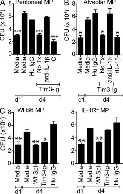
IL-1β is necessary and sufficient to mediate Mtb killing. Mtb-infected WT peritoneal (A) or alveolar (B) macrophages (MP) were cultured either alone or in the presence of 10 µg/ml Tim3-Ig fusion protein with and without 25 µg/ml anti–IL-1β neutralizing antibody or isotype control (IC). No Tx, treatment with Tim3-Ig alone in the absence of neutralizing antibodies. Data in A is representative of 5–11 independent experiments. Data in B is from one experiment. Bars indicate mean ± SEM from three to six replicate cultures. (C) WT C57BL/6J or IL-1R−/− macrophages were infected with H37Rv in parallel. On day 1 (d1), WT splenocytes, Tim3-Ig, or HuIgG (control) were added to the macrophages. CFUs were determined on d1 and day 4 (d4) after infection. Representative data from three independent experiments are shown. CFUs were determined on day 1 and day 4 after infection. Data are from one experiment. Error bars indicate mean ± SEM from three to six replicate cultures. *, P < 0.05; **, P < 0.01; ***, P < 0.001, one-way ANOVA compared with day 4 macrophages alone.

