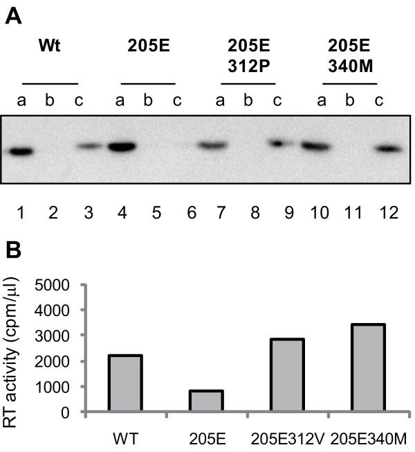
Figure 7.
SIV core stability in vitro. Concentrated SIVmac239 (Wt; lanes 1-3), SIVmac239Gag205E (205E; lanes 4-6), SIVmac239Gag205E312P (205E312P; lanes 7-9), or SIVmac239Gag205E340M (205E340 M; lanes 10-12) was separated into three fractions (top [a], middle [b], and bottom [c]) by ultracentrifugation under gradient sucrose concentrations in the presence of 0.6% Triton X-100. Each fraction was subjected to Western blot analysis to detect SIV CA p27 proteins (A). A representative result from three sets of experiments is shown. The bottom (c) fractions were also subjected to RT assay (B).

