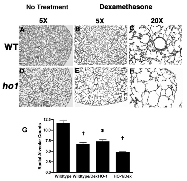
Figure 3.
Dexamethasone treatment exacerbates the alveolar defects in HO-1 mutant mice. A-F: Representative H&E staining of mouse lung sections at P14 at 5X and 20X magnifications. A-C: Wildtype; D-F: HO-1 -/-. A, D: lungs from untreated animals. B, C, E, F: lungs from Dex-treated animals (P3-P14, 0.25 ug/pup/day). In untreated group, lungs from HO-1-/- animals showed simplified, enlarged, and disorganized alveolar structure (A, D). Postnatal Dex treatment in wildtype animals resulted in alveolar simplification and loss of secondary septation (A, and B, C). Dex treatment in HO-1 -/- animals resulted in more dramatic disruption of the alveolar structure with larger alveolar space, thinning of the alveolar wall, and lack of secondary septation. V: pulmonary vasculature. A: airway. Arrowhead indicates the normal secondary septae. Arrows indicate the elongated and thinning of the alveolar wall. G: Quantification of alveolar development by RAC of the lung sections at P14. * P < 0.05 vs. wildtype, † P < 0.05 vs. untreated group of same genotype. n = 3-4 for each group.

