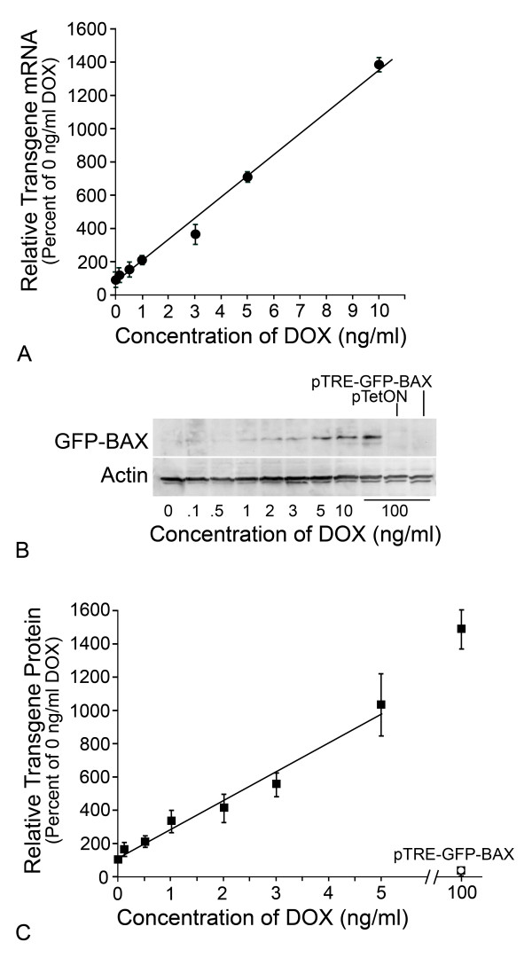
Figure 2.
Titration of GFP-BAX transgene expression in HCT116BAX-/- cells. HCT116BAX-/- cells were co-transfected with pTetON and pTRE-GFP-BAX and exposed to increasing concentrations of DOX in the media. (A) Quantification of transgene transcripts using quantitative (Real-Time) PCR. Data shown were normalized to S16 cDNA in each sample and indicated as a percentaqe of the transcript level at 0 ng/ml DOX (mean ± SEM of triplicate samples from 3 separate experiments used for calculations). Levels of transgene mRNA increased linearly with increasing concentrations of DOX up to 10 ng/ml (R2 = 0.991). (B) Immunoblot of transgene expression. GFP-BAX protein expression increased with increasing concentrations of DOX. Low levels of GFP-BAX were detected in cells treated with 0 ng/ml DOX, but not in controls of each plasmid transfected individually (right hand lanes). ACTIN levels in each lane are shown as a loading control. (C) Quantification of GFP-BAX protein expression. Transgene protein levels were quantified, normalized to the amount of ACTIN present in the same sample, and expressed as the percentage of protein levels detected in 0 ng/ml DOX (mean ± SEM of data collected from 5 independent gels). Similar to transcript levels, increasing concentrations of DOX produced a linear increase in transgene protein expression (between 0 and 5 ng/ml DOX, R2 = 0.975).

