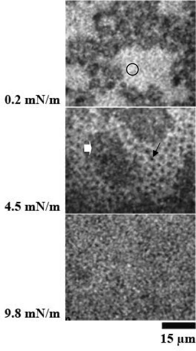
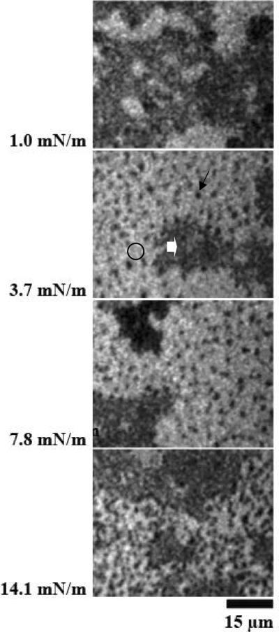
Figure 1.
Image of monolayers of DPPC containing 17% SP-B spread over a subphase of 0.68 μg/ml of porcine SP-A (Panel A) and human SP-A (Panel B). Surface pressures at which images were obtained are given at the sides of the Figure. Three phases in the images were observed, including lipid liquid expanded (LE) phase (bright regions, circles), lipid liquid-condensed (LC) phase (small black domains, arrows), and surface clusters characteristic of SP-A and SP-B complexes (grey regions, block arrows). The morphology of the DPPC monolayers containing SP-B and human SP-A in panel B was similar to that of the lipid monolayers plus SP-B and porcine SP-A in panel A.


