Figure 1.
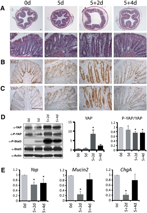
Increased YAP protein levels in regenerating crypts. (A) H&E staining of colon sections from 8-wk-old wild-type mice before (0 d) and after a 5-d DSS treatment (5 d), followed by normal drinking water for 2 d (5 + 2 d) or 4 d (5 + 4 d). (Top row) Low magnification. (Bottom row) High magnification. (B) Ki67 staining. Note the restriction of Ki67-positive proliferating cells to the crypt base in a normal colon, the loss of Ki67-positive cells in 5-d crypts, the expansion of Ki67-positive cells from the crypt base to the whole regenerating crypt in 5 + 2-d colons, and the restoration of Ki67-positive cells to the crypt base in 5 + 4-d colons. (C) YAP staining. In wild-type crypts, YAP was detected in the entire crypt epithelium. Note the dramatic increase of YAP staining in 5 + 2-d crypts. Also note the absence of appreciable YAP nuclear accumulation in control and regenerating crypts. (D) Western blotting analysis. Protein extracts from control and regenerating crypts were probed with the indicated antibodies. Note the increase of YAP, P-YAP, and P-Stat3 in 5 + 2-d and 5 + 4-d crypts. The YAP protein level and P-YAP/YAP ratio were quantified in the graphs to the right. Data are mean ± SD. n = 3. (*) P < 0.01, t-test. (E) Real-time PCR analysis. mRNAs from control and regenerating crypts were analyzed for the expression of the indicated genes. Note the decrease of Yap mRNA in 5 + 2-d and 5 + 4-d crypts. Also note the decreased expression of the goblet cell marker Mucin2 and the enteroendocrine cell marker Chromogranin A (ChgA) in 5 + 2-d crypts, and the recovery of their expression levels in 5 + 4-d crypts. Data are mean ± SD. n = 3. (*) P < 0.01, t-test. Bars, 100 μm.

