Figure 4.
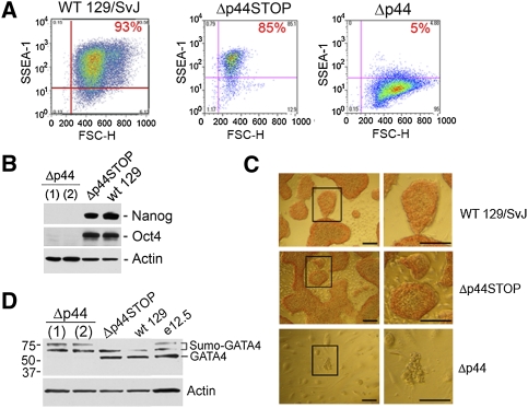
Reduced Δ40p53 expression leads to spontaneous loss of ESC pluripotency. (A) Reduced SSEA-1 expression in ESCs haplosufficient for Δ40p53. FloJo FACS analysis of SSEA-1 staining (Y-axis) plotted against forward scatter (FSC) (X-axis) in wild-type (129/SvJ; left), p53+/Δp44STOP (Δp44STOP; middle), and p53+/Δp44 (Δp44; right) ESCs. (B) Loss of stem cell markers in ESCs with reduced Δ40p53. Western blot analysis of Nanog and Oct4 expression in p53+/Δp44 (Δp44) cells relative to wild-type (129/SvJ) or p53+/Δp44STOP (Δp44STOP) ESCs. Δp44(1) and Δp44(2) represent two unique p53+/Δp44 clones. (C) Decreased AP activity in ESCs with reduced Δ40p53. Histochemical detection of AP activity in p53+/Δp44 (Δp44; bottom), p53+/Δp44STOP (Δp44STOP; middle), and wild-type (129/SvJ; top) ESC colonies. Images are phase-contrast photomicrographs of cells incubated with a substrate that is converted to a red reaction product in the presence of AP. Bar, 100 μm. (D) Altered GATA-4 expression in ESCs with reduced Δ40p53. Western blot analysis of GATA-4 expression in p53+/Δp44 (Δp44), wild-type (129/SvJ), or p53+/Δp44STOP (Δp44STOP) ESCs and embryonic day 12.5 (E12.5) embryonic tissues. High-molecular-weight bands correspond to sumoylated forms of GATA-4, and are described fully in the text.

