
Figure 2.
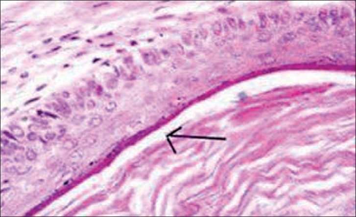
Histopathological findings of fibrous tissue lined in combination with compressed stratified squamous epithelium (arrow), conclusive of epidermoid cyst, (H & E, 40×)
Official websites use .gov
A
.gov website belongs to an official
government organization in the United States.
Secure .gov websites use HTTPS
A lock (
) or https:// means you've safely
connected to the .gov website. Share sensitive
information only on official, secure websites.
Histopathological findings of fibrous tissue lined in combination with compressed stratified squamous epithelium (arrow), conclusive of epidermoid cyst, (H & E, 40×)