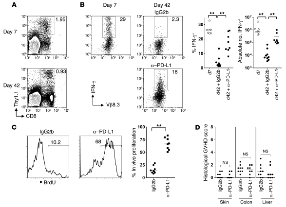
Figure 5. Minor H antigen–specific CD8+ T cells recognizing ubiquitously expressed host antigens are susceptible to exhaustion.
B6 female CD3 cells (8 × 10⁶) and Mh Tg Thy1.1+ CD8+ cells (106) were transferred 1 week after lethal irradiation of B6 male mice and reconstitution with female B6 BM. At 36 and 39 days following T cell transfer, mice were administered 0.2 mg anti–PD-L1 blocking antibody or isotype control by i.p. injection and received BrdU in the drinking water during the same period. (A) Representative contour plots showing frequency of Thy1.1+ CD8+ cells as a percentage of SCs. (B) SCs recovered from chimeras at 7 or 42 days after T cell transfer were stimulated overnight with UTY or irrelevant peptide. Representative dot plots and summary data show IFN-γ production by CD8+ Thy1.1+ Mh T cells. Gates set according to irrelevant peptide. (C) Representative histograms and summary data showing the percentage of Thy1.1+ CD8+ T cells incorporating BrdU. (D) Single-blind scoring of histological GVHD in sections taken from skin, colon, and liver of recipient mice. Data derived from 4 independent experiments. **P < 0.01, Mann-Whitney test.

