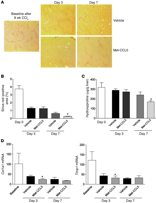
Figure 7. Met-CCL5 accelerates the regression of liver fibrosis in vivo.
(A) C57BL/6 mice were challenged with CCl4 for 8 weeks to establish advanced liver scarring. Three days after the last the last CCl4 injection (at the peak of fibrosis), mice received either Met-CCL5 or vehicle (n = 8/group) and were assessed for fibrosis regression by histology for an overall duration of 7 days. At day 7, the mice that received Met-CCL5 displayed a significantly reduced residual fibrosis compared with the vehicle-treated group (original magnifications, ×40). (B) The difference between the groups is evidenced by a reduced Sirius red–positive area in the Met-CCL5–treated mice (*P < 0.05) and (C) by significantly lower hydroxyproline contents at the same time point during fibrosis regression (*P < 0.05). (D) Functionally, mRNA expression of Col1a1 and Timp1 is already significantly reduced at day 3, after start of Met-CCL5 or vehicle treatment (*P < 0.05).

