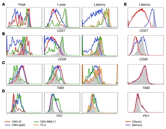
Figure 6. HCMV-specific cells show extensive differentiation, but do not become exhausted.
CD8+ T cells from a patient experiencing primary HCMV response and from a healthy latent virus carrier were stained with tetramers specific for HCMV and EBV epitopes. Shown is 1 representative patient of 3 and 1 representative healthy virus carrier of 4 analyzed. (A) CD27. (B) CD28. (C) TIM3. (D) PD1. (E) Expression on total memory and effector CD8+ T cells. Filled gray histograms denote staining of naive CD8+ T cells.

