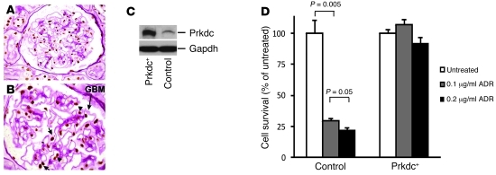
Figure 3. PRKDC is expressed in podocytes, and its overexpression protects against ADR cytotoxicity.
Immunohistochemistry of human kidney with PAS counterstain demonstrates expression of PRKDC (brown staining) in the nuclei of podocytes (short arrows) and endothelial cells (arrowhead) of the glomerulus. The glomerular basement membrane (GBM [long arrow, magenta]) and Bowman’s capsule landmarks allow identification of podocytes by their anatomic location, as podocytes are the only glomerular cell type overlying the glomerular basement membrane in the urinary space (outside the glomerular capillaries). Original magnification, ×600 (A); ×1,000 (B). (C) The Western blot shows an increased level of Prkdc in murine podocyte clone stably overexpressing Prkdc (Prkdc+) as compared with that of control podocytes. The positions of Prkdc and Gapdh (loading control) are indicated by arrows. (D) Comparison of survival among control and overexpressing Prkdc (Prkdc+) podocytes after treatment with 0.1 and 0.2 μg/ml ADR The ADR-treated groups were compared with the untreated control within each cell type.

