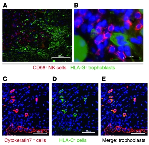
Figure 2. HLA-C is expressed predominately on the EVT in the decidua basalis.
(A and B) Immunofluorescence staining of frozen sections of human first-trimester implantation site showed infiltrating EVTs in sections of decidua basalis identified by staining for HLA-G. EVTs were observed mingling and in close apposition with CD56+ NK cells. (C) Cytokeratin staining identified EVTs and glandular epithelial cells. (D and E) Only EVTs were strongly labelled with the mAb DT9, specific for HLA-C allotypes. There was weak staining of other maternal cells in the decidua identified as CD45+ leukocytes (not shown). Scale bars: 200 μm (A); 30 μm (B); 60 μm (C–E).

