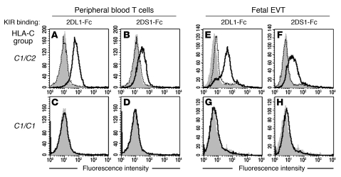
Figure 4. KIR2DS1 and KIR2DL1 bind specifically to C2 molecules on EVTs.
(A–H) Binding of KIR-Fc fusion proteins was measured by flow cytometry to peripheral blood CD3+ T cells (A–D) or HLA-G+ EVTs isolated from placentae of healthy first-trimester pregnancies (E–H). KIR2DL1 and 2DS1 bind cells from C2-positive (A, B, E, and F) but not C2-negative (C, D, G, and H) donors. The indicated KIR-Fc fusion protein histograms are outlined in black and secondary antibody histograms in gray. Pre-incubating with an anti-KIR mAb (HP-3E4) blocked KIR2DS1-Fc and KIR2DL1-Fc binding to both PBLs and trophoblasts (histograms with dotted line).

