
Fig. 2.
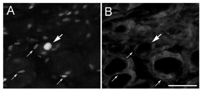
ATF3-IR in satellite cells in the SCG after decentralization. The CST was cut and 48 h later, sections of the SCG were double immunostained for ATF3 (A) and S100 (B). A labeled principal neuron is indicated by the large arrow, and three labeled satellite cells are indicated by the small arrows. Satellite cells are distinguished from Schwann cells by their apposition to principal neurons. Scale bar = 25 μm.