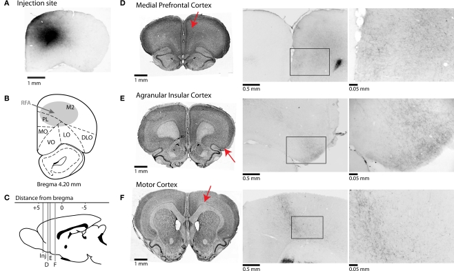
Figure 2.
Afferent projections from RFA to frontal cortical regions. (A) Photomicrograph of injection site from one case stained for BDA. (B) Representative section showing approximate location of injections into the rostral forelimb area (RFA). (C) Mid-sagittal representative section showing AP-coordinates of projections of rostral forelimb area to multiple cortical areas. Red arrows indicate the approximate locations of the sections in the subsequent panels. (D–F) Corticocortical projections of rostral forelimb area. Nissl-stained sections shown on left represent coronal slices to which BDA-stained sections in middle and right sections correspond. Photomicrographs in center are taken at 4× magnification, and at right at 40× magnification. (D) RFA projects to areas of anterior cingulate cortex and prelimbic cortex, and fibers are seen in superficial layers of prelimbic at higher magnification. (E) Connections to agranular insular cortex are seen at high magnification across superficial and deeper layers of cortex. (F) Projections are found in deeper layers of primary motor cortex.

